![Diseases, Symptoms, tcm, [tcmwindow.com]](/uploadFile/adImg/2015/11/11/f5cbfcc0-4df5-4646-9b9a-f316651a0199.jpg)


Computed tomography (CT) scanning, also referred to as, Computed axial tomography (CAT) scanning is a unproblematic, noninvasive and effortless health investigation. It aids doctors to detect and take care of medical conditions. CT scans are more preferred than x-rays, as they provide greater precision and accuracy. CT examinations are swift and undemanding and divulges inner injuries and blood loss, thereby saving lives. Irrespective of any implanted devices, this procedure is effective.
CT imaging utilizes an x-ray apparatus with an attached computer to r
CAT scan
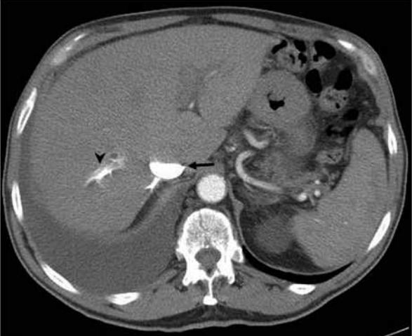
eview the images and a print out can be taken. This method is used to visualize the kidneys, liver, gall bladder, spleen, ovaries, aorta and uterus. Tumors, body changes due to trauma, abnormal anatomy are verified using a CAT scan. All abdominal pains and diseases are easily diagnosed by an abdominal cat scan. All conditions, including Diverticulitis, Kidney Stone, organ injury or body changes due to trauma, obstruction of bowels and appendicitis are detected using a CAT scan.
The CAT scan machine looks like a huge, tapering doughnut. A platform moves towards the central hole of a machine and the patient lies down on this platform. The X-ray tube is installed on a impermanent ring in the region of the hole edge. A display of X-ray detectors is unswervingly placed opposite the X-ray tube. An electrical motor rotates the ring keeping the X-ray tube and the detectors in rotation around the body. Every part of the body is scanned. The control system mobilizes the display place beyond the hole so as to diagnose the next part. The x-ray is recorded in a spiral way. The x-ray intensity is adjusted to optimum power by the computer.
A detailed image is formed by processing and combining all the scan information. Entire body scan is unnecessary. It is a procedure which has no or low risks. A contrast ingredient, oral or rectal, might be of use, during a scan. Commonly used contrast is dilute barium sulfate. Other agents, like, iodinated contrasts are used in cases of injury.
The scanned image is read and interpreted by a trained physician or radiologist. Adverse reactions to intravenous contrast (iodine-based), which lasts a few minutes or hours, includes rashes, warmth feeling and hives. These contrasts highlight the kidneys and blood vessels and make them more perceptible. Anti-histamines provides relief from these symptoms.
Magnetic Resonance Imaging differs from CT Scan. There is no radiation exposure in the former. The MRI paraphernalia comprises of two potent magnets, external and internal.When the negative and positive charged atoms of the body are open to the elements of electromagnetic waves from the MRI equipment, the atoms behave like magnets. The data obtained by the computer is composed, pooled, and manipulated.